
Fluorescein Angiography and Ophthalmic Photography
What?
There are two types of photography that you may encounter when you attend an eye clinic.- Colour photography of the retina, also called the fundus.
- Fluorescein Angiography, an intra-venous dye test that dynamically images the retinal blood vessels.
Where?
In order to take photographs or to examine the retina, dilating drops are placed into the eye which prevent the iris muscle reacting to light and contracting thus giving a clear view of the retina. Once the eyes are dilated and after examination by the Ophthalmologist, you may be sent through for photography to record the retina and sometimes fluorescein angiography as well.Fluorescein Angiography involves the injection of a yellow dye which combines with the protein in the blood and fluoresces, that is, it gives off light of a higher wavelength when exposed to blue light. The bio-microscope camera that is used has the blue filter built in to it, it also has a yellow filter which cuts out any visible light so that the images photographed are of the fluorescence of the dye in the blood vessels and other eye pathology.
Why?
The dye test is done to assess if there is any inflammation in the retina, also how much inflammation there is and where it is located within the retina. This can help the ophthalmologist in assessing the condition and the level and type of treatment required.When?
It is more likely that this test will be done more regularly at the beginning of your clinic visits, primarily for initial diagnosis and after that for assessment of response to treatment. It may also be done if your vision deteriorates from the last visit.How?
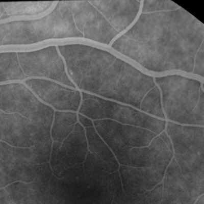
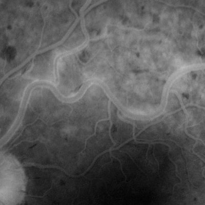
Colour photographs and green photographs are taken first of the retina usually of a 'straight ahead' view which will show the area of sharpest vision (macula). After that a butterfly needle or ventflon (plastic tube) is placed in an arm or hand vein and the dye is injected rapidly over 5 seconds. After around 14 seconds the dye starts to be seen circulating in the blood vessels of the retina and a rapid sequence of flash photos is taken for the first minute or so. After that photos may be taken of the peripheral parts of the retina usually four quadrants; and of the other eye. The last photographs are taken at 5 minutes from the initial dye injection.Shown below are two examples of flourescein angiograms. The first one shows a normal appearance. The dye passes through the blood vessels with no "blockages" and no leakage. The vessels look nice and sharp. Take a look at the second one and you will see the dye "leaking" out of the blood vessels, which look rather fuzzy. this angiogram comes from a patient who has active uveitis.
1. Normal angiogram
2. Angiogram showing active inflammation
 Some people experience a 'funny taste' as the dye goes in, and others
feel slightly sick for a short while, but this usually goes away very
quickly. The dye may make the skin look a little yellow afterwards for
an hour or so, and the urine turns a sort of fluorescent green for a
couple of days, whilst the dye clears the body. Visually many people
say they experience a pink colour cast to everything in the room for a
short while afterwards. Any other shorter term focusing difficulties
and intolerance to bright lights are usually linked with the dilating
drops used, and for this reason it is not recommended to drive
afterwards, indeed extra care also needs to be taken if you are a
pedestrian.
Some people experience a 'funny taste' as the dye goes in, and others
feel slightly sick for a short while, but this usually goes away very
quickly. The dye may make the skin look a little yellow afterwards for
an hour or so, and the urine turns a sort of fluorescent green for a
couple of days, whilst the dye clears the body. Visually many people
say they experience a pink colour cast to everything in the room for a
short while afterwards. Any other shorter term focusing difficulties
and intolerance to bright lights are usually linked with the dilating
drops used, and for this reason it is not recommended to drive
afterwards, indeed extra care also needs to be taken if you are a
pedestrian.
Occasionally an allergic reaction may occur, this can range from sneezing or slight itching and tingling of skin, sometimes just around the injection site, to a massive reaction similar to that of some individuals to a bee sting. This kind of reaction is extremely rare and all drugs and equipment needed to treat any degree of reaction are situated in the camera room. In Aberdeen alone around 2000 uneventful Fluorescein Angiographs take place each year and there are many other centres also performing this test on a routine basis.