
Glossary of Terms
This section aims to cover a lot of the terms we may come across, when reading or talking about uveitis. It will, hopefully, give clear, correct explanations instead of complicated medical definitions.The group aims to help people understand their condition so that they can deal with it most effectively and positively. One result, hopefully, of a better understanding, is to make discussing things with our doctors easier and more useful to both doctor and patient.
When talking or reading about our condition it is likely that all sorts of terms will be used which may appear familiar, like "iris" and "inflammation" or fairly unfamiliar e.g. "synechiae". Although a word like "iris" will seem like an everyday expression, do we really know what it means? The aim of explaining these terms is to help understand our doctors and to aid our discussions with them.
The list is not exhaustive (yet) and may well be added to. If there any terms you have come across that are not here, then please ask for a "request".
The glossary does not include explanations of all the different names of types of uveitis or medical conditions associated with it. These are dealt with in fact sheets or from specific requests for information. They may well be included in a separate glossary in future.
A B C D
E F G H
I J K L
M N O P
Q R S T
U V W X
Y Z
A
topAmsler Grid: A chart made up of grid lines and a central dot. It is very useful to detect and monitor problems with the macula, or central vision.
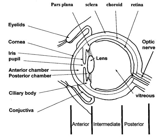
Anterior is simply a word for "front" and in the eye is taken to describe the part of the eye from the lens, forwards to the cornea. (see diagram).
Anterior chamber: the part of the eye in between the iris and the cornea(see Diag. Page 5). It is filled with a viscous fluid, the aqueous
Anterior uveitis: an inflammation of any part of the anterior uvea which is made up of the iris and the ciliary body
Antigen: Something potentially capable of causing an immune response.
Antibodies: are specialised proteins produced by white blood cells that circulate in the blood seeking and attaching to foreign proteins, microorganisms or toxins in order to neutralise them. They are part of the immune system.
Aqueous: Fresh fluid called aqueous is constantly produced by an organ called the ciliary body located behind the iris. The fluid then circulates from behind the iris through the pupil, moves through the anterior chamber and finally exits the eye through a drainage mechanism called the trabecular meshwork. (see diagram).
Autoimmune diseases: Illnesses which occur when the body tissues are attacked by its own immune system.
B
topC
topCentral Vision involves the macula which is in the centre of the retina. As we look "straight ahead" to perform such tasks as reading, recognising faces and doing any work requiring fine, sharp vision, then the light is focused onto our macula. There, millions of cells change the light into nerve signals that tell the brain what we are seeing. This is called our central vision.
Choroid is a part of the uvea. It runs from the ciliary body around the back of the eye between the retina and the sclera. (see diag. p. 4). It is pigmented and is mainly made up of blood vessels which supply the retina.
Chronic: comes from the Greek "chronos" meaning time. In medicine, It means lasting a long time. A chronic condition is generally taken as one lasting 3 months or more, In ancient Greece, the "father of medicine" Hippocrates distinguished diseases that were acute (abrupt, sharp and brief) from those that were chronic.
Ciliary Body is a circular, pigmented part of the uvea. It produces the clear fluid, the aqueous which fills the part of the eye in front of the lens. It also helps change the eye's focus via muscles attached to the lens.
Conjunctiva: a thin, moist layer which lines the inside of the eyelids and part of the outer surface of the eye. (see diag.).
Conjunctivitis is an inflammation of the conjunctiva, and is a common cause of the so called "red-eye".
Cornea: The clear front window of the eye. The cornea transmits and focuses light into the eye. Inflammation of the cornea is called keratitis
D
topE
topF
topFluorescein Angiography: involves the injection of a dye into the arm, followed by a series of photographs which captures the fluorescent dye as it passes through the blood vessels at the back of the eye. It is a useful way to assess inflammation and problems around the macula.
Fundus Although Latin scholars will know that this is the word for the bottom, in medicine the fundus refers to the base of an organ. As far as the eye goes, it describes what the ophthalmologist sees at the back of the eye by looking through the pupil, I.e. the retina, optic nerve and the macula.
G
topH
topI
topImmune System: A complex system that is responsible for distinguishing us from everything foreign to us, and for protecting us against infections and foreign substances.
Immunosuppression: Lowering the immune response, for example, with immunosuppressant drugs, to treat auto-immune diseases.
Inflammation: The reaction of living tissues, especially the small blood vessels and the blood cells within them, to injury. The term "injury" may include trauma, infection or over activity of the immune system. (see also "The treatment of uveitis", newsletter no.1 Oct.98 ).
Intermediate uveitis: describes the region of the eye affected most by the inflammation. See also pars planitis.
Intraocular lens(IOL): When a clouded lens, (cataract) is removed, it is usually replaced by a plastic intraocular lens (IOL). Cataract surgery is generally more complicated in people with uveitis and there are situations where this type of lens may not be recommended by the doctors.
Intraocular pressure(IOP): The ciliary body produces a fluid, the aqueous. One of the functions of this is to keep a pressure inside the eye to keep its shape, a bit like air in a balloon. The aqueous enters the posterior chamber, passes through the pupil into the anterior chamber (see diagram) and then drains through a sieve-like structure and then though a canal into the blood stream. If the amount of fluid getting produced and the ability for it to be drained away gets "out of balance" then the intraocular pressure will be affected. A rise in the IOP may cause glaucoma.
Iris is the most anterior part of the uvea. It is a coloured ring of tissue, containing muscle fibres which regulate the amount of light entering the eye by adjusting the size of the pupil.
Iritis: inflammation of the iris. Strictly speaking, it is a term which is wrongly used in place of the better term, anterior uveitis, because there is also, often, inflammation of the ciliary body. (iridocyclitis). If you have been told you have "iritis", you will then have anterior uveitis.
"itis" Any word ending in "itis" is describing an inflammation. Uveitis is an inflammation of the uvea and arthritis an inflalmmation of the joints. This is another example of the use of Greek terms in medicine.
J
topK
topKeratitic precipitates(KP's): Accumulations of inflammatory cells that "stick" to the inside of the cornea, seen at an eye examination. They are useful in the diagnosis of anterior uveitis.
L
topLow vision: When ordinary eyeglasses are unable to bring a patient's sight up to normal sharpness and normal daily tasks are affected.
Low vision aids: any aid ranging from magnifiers, computer technology and special lighting which can enable people with low vision to improve their vision.
M
topMacular oedema is an important complication of uveitis. It can occur in all types of uveitis. The macula is affected by a build up of fluid (oedema) It left untreated it can be a significant cause of vision loss.
Macular vision: see central vision
Mydriatic: A drug that dilates (widens) the pupil. They may be short or long acting. A mydriatic works by "paralysing" the muscles of the iris and ciliary body. (It is the movement of these inflammed muscles that causes the pain in anterior uveitis). Apart from treating the symptoms of anterior uveitis and preventing complications, they are also used to widen the pupil to allow the Ophthalmologist to examine the eye.
N
topO
topOptic Nerve connects the eye to the brain. It carries the impulses created at the retina to the brain. In a sense the optic nerve is an extension of the brain.
P
topPhotophobia: Painful, oversensitivity to light. This is often a symptom of anterior uveitis.
Posterior chamber: The space within the eye between the back of the iris and the lens(see diagram). It is filled with a fluid, the aqueous. Take care not to confuse this space with the larger space behind the lens containing the vitrieous. (see diagram).
Posterior synechiae: see synechiae
Pupil is the dark aperture in the iris that lets light into the eye. The size of the pupil is changed by the muscles in the iris. It is widened, (dilated) to let more light in and narrowed (constricted) to let less light through.
Posterior uveitis: the inflammation is located at the back of the eye.
Pupil dilation Drops are placed in the eyes before an eye examination to dilate, or widen the pupil so that the Ophthalmologist can look clearly at the back of the eye, the fundus. This will cause a temporary blurring of vision and make the eyes very sensitive to light for a while. Pupil dilation will also occur in the treatment of anterior uveitis by mydriatic drops.
Q
topR
topRefraction: the bending of light that takes place within the human eye. Refractive errors like short sightedness can be corrected by wearing glasses.
Retina: the nerve layer that lines the back of the eye, senses light and creates impulses that travel through the optic nerve to the brain.
S
topScotoma: A blind or partially blind area in the visual field.
Synechiae: These occur when, due to inflammation, the iris "sticks to the lens just behind it. (posterior synechiae). They are an important complication of anterior uveitis, often leading to secondary glaucoma. They may also make examination of the eye and cataract surgery more difficult. Synechiae may also be described as "adhesions".
T
topU
topUveitis an inflammation of any part of the uvea.
V
topVisual field: The entire area which can be seen without shifting the gaze. It is a measure of the peripheral vision. There are different instruments to determine the visual field in the eye clinic.
Vitrectomy: Removal of the gel (vitreous) from the centre of the eye. This may be done because it has blood and scar tissue in it that blocks sight. An eye surgeon replaces the clouded gel with a clear fluid.
Vitreous: A clear, jelly-like substance that fills the middle of the eye, in between the lens and the retina.(see diagram).